Diagnostics
3D CT and dental X-rays
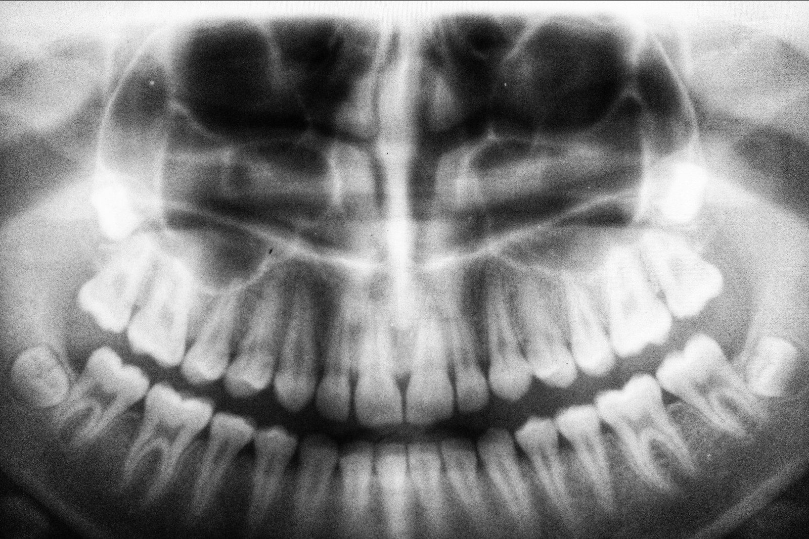
An X-ray shows what cannot be seen at an examination: the roots, the canals, the bone and hidden areas of inflammation. Which type of imaging is needed is decided by the doctor, depending on the task.
- from 60 000 UZSX-ray of a tooth
- Three typesX-ray of a tooth, panoramic X-ray and 3D tomography
- As prescribedthe doctor chooses the type of imaging to suit the treatment task
01 / What it is
Why X-rays are needed
At an examination the doctor sees only the crowns of the teeth and the gum. The condition of the roots and canals, caries under a filling or between the teeth, inflammation at the root apex, the bone level — all of this is shown by an X-ray. The doctor uses it to refine the diagnosis, plan the treatment and monitor its result.
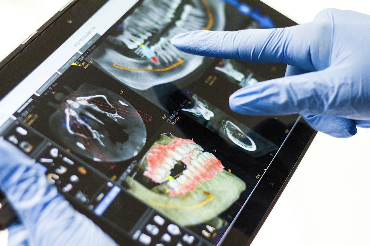
Ordinary X-rays are flat: three-dimensional structures are superimposed on one another. Computed tomography gives a three-dimensional image that can be viewed slice by slice and in different planes. At the same time, the doctor prescribes the type of imaging that is sufficient for the specific task and does not refer you for an X-ray without an indication.
Who it is for
- Implant placement or a sinus lift is planned
- A wisdom tooth or an impacted tooth is to be extracted
- Root canals need to be treated or retreated
- The tooth hurts, but the cause is not visible at an examination
- There is gum disease and the condition of the bone needs to be assessed
- A plan for prosthetic or comprehensive treatment is being drawn up
If you are pregnant or may be pregnant, be sure to tell the doctor before the imaging: the doctor will decide whether it can be postponed or replaced.
02 / Stages
How treatment goes
-
Stage 01
Referral
At the examination the doctor determines which X-ray is needed and which area to image. If you have recent X-rays, bring them with you.
-
Stage 02
Preparation
No special preparation is required. Before a panoramic X-ray and tomography you will be asked to remove metal jewellery, glasses and removable dentures in the head and neck area.
-
Stage 03
Imaging
A member of the clinic staff helps you take the correct position. You need to stay still during the imaging: the sharpness of the image depends on it.
-
Stage 04
Interpretation
The doctor studies the image, shows you the findings on the screen and explains what they mean.
-
Stage 05
Treatment plan
Based on the examination and the X-ray, the doctor proposes treatment options and answers your questions.
03 / Prices
How much it costs
The doctor's consultation is paid for separately. The type of imaging is determined by the doctor after an examination.
Full price list04 / Questions
Frequently asked questions
How does a CT scan differ from a panoramic X-ray?
A panoramic X-ray is a flat overview image of both jaws. Computed tomography is three-dimensional: the doctor sees the jaw slice by slice and in different planes, and can assess the bone thickness and the position of the nerves and sinuses. Tomography does not replace a panoramic X-ray in every case — it is prescribed when a flat image is not enough.
Are dental X-rays harmful?
Any X-ray examination involves a radiation dose, so it is carried out only when indicated, when the diagnostic benefit outweighs the possible risk. The doctor chooses the type of image that is sufficient for the task and does not prescribe examinations “just in case”. If you have any doubts, discuss them with the doctor before the imaging.
Can X-rays be taken during pregnancy?
The doctor must be told about pregnancy in advance. Routine examinations are usually postponed, and in the case of acute pain or inflammation the doctor assesses whether it is possible to manage without an X-ray and makes the decision individually.
Are X-rays from another clinic acceptable?
Bring them with you — the doctor will take a look. If the X-ray is recent, of good quality and shows the required area, the examination may not need to be repeated. A new X-ray will be needed if the situation has changed since it was taken or if the image is not sufficient for planning.
Start with a consultation
Consultation and examination by a doctor — 150 000 UZS. The doctor will draw up a treatment plan and tell you the exact cost.